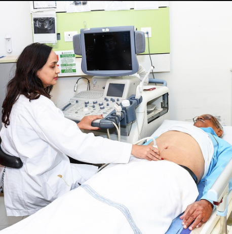
Ultrasound is one of the most commonly used medical imaging techniques for examining internal organs, monitoring pregnancy, and identifying certain health conditions. It uses high-frequency sound waves to create images of structures inside the body without using ionising radiation. Doctors may recommend an ultrasound when they need to investigate pain, swelling, pregnancy-related concerns, or abnormalities detected during a physical examination.
There are many types of ultrasound tests, and each serves a different medical purpose. Some examine the abdomen, kidneys, heart, or thyroid, while others help doctors monitor a developing baby or assess blood flow through the body’s blood vessels.
There is no single fixed number of ultrasound tests because different examinations can be classified by the body part examined, the technology used, or the clinical purpose. However, several major types are routinely performed in hospitals and diagnostic centres.
This article explains the main types of ultrasound tests, their uses, how they are performed, and what patients should know before an appointment.
How Many Types of Ultrasound Tests Are There?
There are more than 10 commonly performed categories of ultrasound examinations, with many additional specialised scans available. The exact number depends on how the examinations are classified.
For example, an abdominal ultrasound and a pelvic ultrasound examine different body regions. Doppler ultrasound evaluates blood flow, while echocardiography examines the heart. Pregnancy ultrasound includes several examinations performed at different stages of pregnancy.
The most common types include:
- Abdominal ultrasound
- Pelvic ultrasound
- Obstetric or pregnancy ultrasound
- Transvaginal ultrasound
- Breast ultrasound
- Thyroid ultrasound
- Kidney and urinary tract ultrasound
- Scrotal or testicular ultrasound
- Doppler ultrasound
- Echocardiography
- Musculoskeletal ultrasound
- Carotid ultrasound
- Transrectal ultrasound
- Neonatal cranial ultrasound
- Ultrasound-guided procedures
Not every patient needs all these examinations. A doctor recommends a particular scan based on symptoms, medical history, and the area that needs investigation.
1. Abdominal Ultrasound
An abdominal ultrasound examines organs inside the abdomen. These commonly include the liver, gallbladder, pancreas, spleen, kidneys, and major abdominal blood vessels, depending on the type of examination requested.
Doctors may recommend this scan when a patient experiences abdominal pain, swelling, or unexplained discomfort. It can help identify gallstones, some liver abnormalities, kidney stones, fluid collections, and certain organ enlargement.
The procedure is generally painless. A technician or radiologist applies a gel to the skin and moves a handheld device called a transducer over the abdomen. Sound waves create images that can be reviewed by the radiologist.
Some abdominal ultrasound examinations require fasting for several hours, particularly when the gallbladder needs to be examined. Always follow the diagnostic centre’s instructions.
2. Pelvic Ultrasound
A pelvic ultrasound examines organs in the lower abdomen and pelvis. In women, it may assess the uterus, ovaries, and surrounding structures. In men, it may examine the bladder and prostate region, depending on the type of scan.
Doctors may request a pelvic ultrasound to investigate pelvic pain, abnormal bleeding, menstrual problems, suspected ovarian cysts, fibroids, or certain fertility-related concerns.
The scan can be performed externally through the lower abdomen. A full bladder may be required to improve visibility, so patients should follow the instructions provided before the appointment.
In some cases, the doctor may recommend a transvaginal ultrasound for more detailed imaging of the uterus and ovaries.
3. Obstetric or Pregnancy Ultrasound
An obstetric ultrasound is performed during pregnancy to assess the developing baby and monitor the pregnancy.
Depending on the stage of pregnancy, the examination may help confirm the pregnancy’s location, estimate gestational age, assess fetal growth, examine the baby’s anatomy, and evaluate the placenta and amniotic fluid.
Common pregnancy scans include:
Early pregnancy ultrasound: Helps establish the location of the pregnancy and assess early development when clinically appropriate.
Dating scan: Estimates how far along the pregnancy is and helps calculate the expected delivery date.
Nuchal translucency scan: Performed during a specific period in early pregnancy as part of screening for certain chromosomal conditions.
Anomaly scan: Usually performed around 18–22 weeks of pregnancy to examine the baby’s anatomy.
Growth scan: Assesses the baby’s growth and other features when clinically indicated, often later in pregnancy.
The timing and number of scans depend on local clinical guidelines, the stage of pregnancy, and individual medical needs. Ultrasound does not detect every possible fetal condition.
4. Transvaginal Ultrasound
A transvaginal ultrasound provides detailed images of the uterus, ovaries, cervix, and surrounding pelvic structures. It is commonly used in gynaecology and early pregnancy assessment.
Instead of moving the transducer over the abdomen, a specially designed probe is gently inserted into the vagina. The probe is covered with a protective covering and lubricating gel.
Doctors may recommend this examination to investigate pelvic pain, abnormal uterine bleeding, ovarian cysts, or certain early pregnancy concerns.
It may provide clearer images than an abdominal scan because the probe is positioned closer to the pelvic organs. Patients can ask the healthcare professional to explain the procedure beforehand, and the examination should be conducted with appropriate consent and privacy.
5. Breast Ultrasound
A breast ultrasound uses sound waves to examine breast tissue. It may be recommended when a person has a breast lump, pain, swelling, or an abnormal finding on another examination.
The scan can help distinguish a fluid-filled cyst from a solid lump and may guide further investigation. However, ultrasound cannot identify every type of breast cancer and does not replace mammography when mammography is indicated.
Doctors may recommend ultrasound alongside a clinical breast examination or other imaging tests, depending on the patient’s age, symptoms, and medical history.
6. Thyroid Ultrasound
A thyroid ultrasound examines the thyroid gland, which is located at the front of the neck. The thyroid produces hormones that help regulate metabolism, energy use, and other body functions.
Doctors may request this scan when they notice a thyroid lump, enlargement, or abnormality during a physical examination. It can help identify thyroid nodules, cysts, and changes in the gland’s structure.
A thyroid ultrasound is painless and usually does not require fasting. The patient lies on an examination table while a transducer is moved gently over the neck.
The scan provides information about the gland’s appearance, but it does not directly measure thyroid hormone levels. Blood tests may also be needed to assess thyroid function.
7. Kidney and Urinary Tract Ultrasound
A kidney and urinary tract ultrasound examines the kidneys and may include the bladder and other parts of the urinary system.
Doctors may recommend this examination for suspected kidney stones, urinary obstruction, recurrent urinary tract infections in selected cases, blood in the urine, or changes in kidney function.
The scan can identify some structural abnormalities, kidney enlargement, and urine retention in the bladder. However, not every kidney stone or urinary tract condition can be detected by ultrasound.
Depending on the requested examination, the diagnostic centre may ask the patient to drink water and arrive with a full bladder.
8. Scrotal or Testicular Ultrasound
A scrotal ultrasound examines the testicles and surrounding tissues. It can help investigate testicular pain, swelling, lumps, injuries, and certain fertility-related concerns.
Doppler imaging may be included to assess blood flow within the testicles. This is especially important when doctors suspect testicular torsion, a condition in which the spermatic cord twists and can cut off blood supply.
Sudden, severe testicular pain is a medical emergency. Anyone experiencing it should seek emergency medical attention immediately rather than delaying care to arrange a routine ultrasound.
9. Doppler Ultrasound
Doppler ultrasound evaluates the movement of blood through blood vessels. It uses changes in sound waves to estimate the direction and speed of blood flow.
Doctors may recommend Doppler examinations to investigate suspected blood clots, narrowed arteries, circulation problems, or abnormalities in blood flow.
Different Doppler studies examine different areas of the body, including the legs, arms, neck, abdomen, and blood vessels supplying a developing baby.
For example, a venous Doppler ultrasound may be used when a blood clot in a leg vein is suspected. An arterial Doppler may assess blood flow through arteries affected by narrowing.
The preparation depends on which blood vessels are being examined.
10. Echocardiography
Echocardiography, commonly called an echo, is an ultrasound examination of the heart. It produces moving images that allow healthcare professionals to evaluate the heart’s structure and function.
An echocardiogram can assess the heart chambers, valves, pumping function, and the movement of blood through the heart.
Doctors may recommend it when a person has symptoms such as unexplained breathlessness, certain heart murmurs, suspected heart failure, or other concerns about heart function.
The most common type is transthoracic echocardiography, in which the transducer is placed on the chest. Other specialised types, including transoesophageal echocardiography, use different approaches and may require additional preparation.
An echocardiogram is different from an electrocardiogram (ECG). An ECG records the heart’s electrical activity, whereas an echocardiogram uses ultrasound to create images of the heart.
11. Musculoskeletal Ultrasound
Musculoskeletal ultrasound examines muscles, tendons, ligaments, joints, and certain soft tissues.
Doctors may use it to investigate sports injuries, tendon inflammation, fluid around a joint, muscle tears, or some soft-tissue lumps.
The scan can also show how certain structures move during movement, which may help assess particular injuries.
Musculoskeletal ultrasound does not replace every other imaging method. X-rays or MRI may be required when doctors need to examine bones or deeper structures in greater detail.
12. Carotid Ultrasound
A carotid ultrasound examines the carotid arteries in the neck, which carry blood to the brain. Doppler techniques are often used to assess blood flow and identify narrowing caused by plaque.
Doctors may request the examination in selected patients when there is a clinical reason to investigate carotid artery disease.
The scan is non-invasive and usually takes place with the patient lying on an examination table. A transducer is moved gently along the sides of the neck.
The test can help assess certain circulation problems, but it is not recommended as a routine screening test for every healthy person without symptoms or specific risk factors.
13. Transrectal Ultrasound
Transrectal ultrasound uses a specialised probe inserted into the rectum to obtain images of nearby structures, particularly the prostate gland.
It may be used in selected investigations of prostate conditions and to guide certain procedures, including some prostate biopsies.
Preparation varies according to the examination and whether a procedure is planned. The healthcare team should explain what to expect and provide instructions beforehand.
14. Neonatal Cranial Ultrasound
A neonatal cranial ultrasound examines the brain of certain newborn babies, particularly premature infants or babies who require evaluation for specific complications.
The scan is usually performed through the fontanelle, the soft spot in a baby’s skull, which allows sound waves to pass through before the skull bones have fully closed.
Doctors may use this examination to look for certain brain haemorrhages or other structural abnormalities in infants at increased risk.
It is painless and does not involve ionising radiation.
15. Ultrasound-Guided Procedures
Ultrasound is also used to guide medical procedures. Rather than serving only as a diagnostic scan, it helps a clinician see internal structures while performing a procedure.
Examples include guiding a needle during a biopsy, helping drain certain fluid collections, and assisting with the placement of some medical lines.
Using real-time imaging can help the clinician guide instruments toward the intended area while avoiding nearby structures.
The preparation, risks, and recovery depend on the specific procedure being performed.
Is Ultrasound Safe?
Ultrasound is generally considered safe when performed by trained professionals for appropriate medical purposes. It uses sound waves rather than ionising radiation, which distinguishes it from X-rays and CT scans.
However, the examination should be performed when medically appropriate and interpreted by a qualified healthcare professional. Pregnancy scans should be used for clinical purposes rather than unnecessary or prolonged imaging.
Ultrasound also has limitations. Sound waves may not provide clear images of every structure, and the result depends on the body part being examined, the equipment, and the patient’s circumstances.
Sometimes, a doctor may recommend another imaging test if ultrasound does not provide enough information.
How Should You Prepare for an Ultrasound Test?
Preparation depends on the type of ultrasound you need.
- Abdominal ultrasound: You may be asked to fast for several hours.
- Pelvic ultrasound: You may need to drink water and arrive with a full bladder.
- Pregnancy ultrasound: Preparation depends on the stage of pregnancy and the scan type.
- Thyroid or breast ultrasound: Usually requires little or no special preparation.
- Kidney and bladder ultrasound: A full bladder may be needed for some examinations.
- Doppler ultrasound: Instructions depend on the blood vessels being assessed.
Always follow the diagnostic centre’s instructions. If you take regular medicines or have a medical condition that affects fasting or fluid intake, ask your healthcare provider how to prepare safely.
Frequently Asked Questions
1. How many types of ultrasound tests are available?
There are more than 10 common categories of ultrasound examinations, with many specialised variations. The exact number depends on how tests are classified.
2. Which ultrasound is used during pregnancy?
Obstetric ultrasound is used to assess pregnancy. Depending on the stage and medical circumstances, it may include dating, anomaly, growth, or other specialised scans.
3. Is an ultrasound test painful?
Most external ultrasound examinations are painless. Some internal examinations or ultrasound-guided procedures may cause temporary discomfort.
4. Does an ultrasound require fasting?
Not always. Some abdominal ultrasounds require fasting, while thyroid and many other scans usually do not. Follow the instructions provided for your specific examination.
5. How long does an ultrasound take?
Many routine ultrasound examinations take approximately 15–30 minutes, although complex scans and specialised procedures may take longer.
6. Can ultrasound detect all diseases?
No. Ultrasound can identify many abnormalities, but it cannot diagnose every condition. Depending on the findings, a doctor may recommend blood tests, CT, MRI, or other investigations.
Conclusion
Ultrasound is a valuable medical imaging technique used to examine organs, monitor pregnancy, assess blood flow, investigate injuries, and guide certain medical procedures. There are more than 10 common categories, including abdominal, pelvic, pregnancy, breast, thyroid, kidney, Doppler, and heart ultrasound examinations.
Each type serves a specific purpose, so the most suitable examination depends on the patient’s symptoms and the doctor’s assessment. Preparation also varies: some scans require fasting or a full bladder, while others need little preparation.
If your doctor has recommended an ultrasound, confirm the exact scan name, cost, preparation requirements, and expected reporting time with the diagnostic centre. Discuss the results with your healthcare professional to understand what they mean for your health and whether further testing is necessary.